Management of Perforated Duodenal Diverticulum: Case Report
Ali Kimyaghalam MD, Kyle Phillips MD, James Smith MD, Prajeet Kaza MSIV
Citation : Kimyaghalam A, Phillips K, Smith J, Kaza P. Management of Perforated Duodenal Diverticulum: Case Report. Asclepius Med Case Rep 2018;1(1):1-3.
It has been reported that duodenal diverticula are common but perforated duodenal diverticulum is a rare entity. At this time, there is no standardized management for perforated duodenal diverticulum. In these cases, patients usually complain of vague abdominal pain and computed tomography scan is mostly ordered used as an important diagnostic method. Diagnosis and the severity of the disease need to be assessed before any intervention. Essentially, the perforated small bowel is treated with surgical intervention. However, conservative treatment with broad-spectrum antibiotics and strict nil per os has been offered in the past for stable patients. Below, we report one case of perforated duodenal diverticulum that we managed with conservative treatment.
Duodenal diverticulum, abdominal pain, perforation
INTRODUCTION
Duodenal diverticula are common and usually asymptomatic, with prevalence at autopsy of 22% [1]. The duodenal diverticula are either congenital or acquired. Acquired duodenal diverticula are the most common and they represent pulsion diverticula due to a protrusion of mucosa, muscularis mucosa, and submucosa through a wall weakness, formed of the papillae of one of those layers and due to this fact, the periampullary area is the most common site for this pathology [1,2]. Complications of the duodenal diverticula include diverticulitis, perforation, digestive bleeding, distension or bile duct obstruction, and perforation of the diverticulum will need a surgical management due to its high mortality rate [3,4]. If surgery is considered, surgical management will be to perform a diverticulectomy and retroperitoneal drainage. We will review one case report of perforated duodenal diverticulum managed conservatively in our hospital.
CASE REPORT
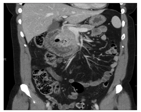
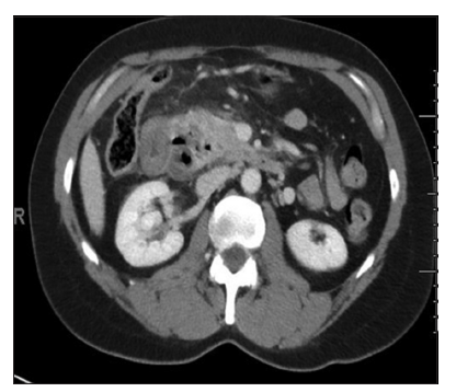
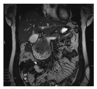
A 44-year-old female presented to the hospital complaining of right upper quadrant dull, achy abdominal pain. Patient had lunch in a restaurant and started having right upper quadrant abdominal pain couple of hours after having lunch. The pain is associated with nausea and bilious emesis, and anorexia. The emesis subsided but the right upper quadrant abdominal pain persisted that made a her come to our emergency department. She denies radiation. She complains of subjective chills and denies fever. Denies history of nonsteroidal antiinflammatory drugs use, denies prior episodes. She had sips of water throughout the day and has lunch last night. She does not recall when she passed gas but had a normal bowel movement 2 days ago. She underwent abdominal ultrasound that showed no abnormality. She subsequently underwent computed tomography (CT) a/p which showed thickened edematous proximal duodenum with 3 cm medial air-fluid collection suggesting for adjacent abscess between duodenum and head of pancreas [Figures 1-3]. She was monitored clinically and was managed conservatively. On hospital day 1, she underwent upper gastrointestinal series that showed edema around the second and third portion of duodenum. During the hospital day 2, her abdominal pain was improving. She underwent magnetic resonance imaging of the abdomen which again showed unchanged appearance of edematous duodenal C-sweep with medial 3 cm abscess and adjacent soft tissue edema/fluid. She was stable to be discharged home. She was advised to have clear liquid diet at the time of discharge. She was advised to follow-up with us in 1 week.
DISCUSSION
The most frequent location for diverticula is the duodenum followed by the jejunum or ileum. They are found to have a difference compared to the colonic diverticula; they are not usually a site of inflammation because of the more sterile and liquid content of the duodenum [8]. Nevertheless, 5% of patient can develop symptoms of acute diverticulitis complicated by fistula, abscess formation, or obstruction of common bile duct [3,8]. There has been < 200 cases of perforated duodenal diverticulum reported in the literature. Most patients present with the right upper quadrant abdominal pain, nausea, and vomiting due to the most perforations being in the retroperitoneum. In our case, the radiologist reported that both CT findings were compatible with duodenal diverticular perforation. Because the disease is rare, there is no standard management protocol for perforated duodenal diverticulum. In general, the surgical approach was considered the treatment of choice. Several technical options are available, ranging from local excision to the Whipple procedure, depending on the location of the duodenal diverticulum and the inflammatory status [3,5]. Our case was treated with conservative management including broad-spectrum antibiotics treatment. Laparoscopic diverticulectomy has also been reported to reduce trauma from laparotomy and achieve an early recovery with minimally invasive surgery [12]. Several recent cases were treated with bowel rest, nasogastric tube and antibiotics, with encouraging results in selected patients [7,9,13]. Conservative treatment may be useful in a patient of advanced age or in a patient with multiple medical comorbidities, provided symptoms are mild and there is no evidence of sepsis. In our case, the patient was stable at the time of admission and stayed hemodynamically stable throughout the admission. Thorson et al.[9] reviewed 40 studies producing 61 cases of perforated duodenal diverticulum from 1989 to 2011. They observed a decrease in mortality and morbidity of this disease for decades because of improved perioperative care, development of broad-spectrum antibiotics, advances in diagnostic tests, and/or increased awareness of this rare disease. Of the 61 cases, 11 patients were managed without surgery, and only one complication was reported, an intra-abdominal abscess treated successfully with percutaneous drainage [6, 10, 11] .
In conclusion, since perforated duodenal diverticulum is a rare condition and symptoms often mimic other intra-abdominal processes, a precise diagnosis is important to manage this disease. To diagnosis this condition appropriately, CT abdomen pelvis can be obtained. The clinical presentation should be considered a guide to management. In a clinically unstable patient, surgical intervention is needed. However, non-surgical treatment should be considered in patients who present with mild symptoms like in our case [14-16].
REFERENCES
- Yin WY, Chen HT, Huang SM, Lin HH, Chang TM. Clinical analysis and literature review of massive duodenal diverticular bleeding. World J Surg 2001;25:848-55.
- Oukachbi N, Brouzes S. Management of complicated duodenal diverticula. J Visc Surg 2013;150:173-9.
- Schnueriger B, Vorburger SA, Banz VM, Schoepfer AM, Candinas D. Diagnosis and management of the symptomatic duodenal diverticulum: A case series and a short review of the literature. J Gastrointest Surg 2008;12:1571-6.
- De Perrot T, Poletti PA, Becker CD, Platon A. The complicated duodenal diverticulum: Retrospective analysis of 11 cases.Clin Imaging 2012;36:287-94.
- Rossetti A, Christian BN, Pascal B, Stephane D, Philippe M. Perforated duodenal diverticulum, a rare complication of a com- mon pathology: A seven-patient case series. World J Gastrointest Surg 2013;5:47-50.
- Miller G, Mueller C, Yim D. Perforated duodenal diverticulitis: A report of three cases. Dig Surg 2005;22:198-202.
- Martinez-Cecilia D, Arjona-Sanchez A, Gomez-Alvarez M, Torres-Tordera E, Luque-Molina A, Valentí-Azcárate V, et al. Conservative management of perforated duodenal diverticulum: A case report and review of the literature. World J Gastroenterol 2008;14:1949-51.
- Coulier B, Maldague P, Bourgeois A, Broze B. Diverticulitis of the small bowel: CT diagnosis. Abdom Imaging 2007;32:228-33.
- Thorson CM, Ruiz PS, Roeder RA, Sleeman D, Casillas VJ. The perforated duodenal diverticulum. Arch Surg 2012;147:81-8.
- Volchok J, Massimi T, Wilkins S, Curletti E. Duodenal divertic-ulum: Case report of a perforated extraluminal diverticulum con-taining ectopic pancreatic tissue. Arch Surg 2009;144:188-90.
- Favre-Rizzo J, López-Tomassetti-Fernández E, Ceballos- Esparragón J, Hernández-Hernández JR. Duodenal diverticulum perforated by foreign body. Rev Esp Enferm Dig 2013;105:368-9.
- Lee HH, Hong JY, Oh SN, Jeon HM, Park CH, Song KY. Laparoscopic diverticulectomy for a perforated duodenal diverticulum: A case report. J Laparoendosc Adv Surg Tech A 2010;20:757-60.
- Ames JT, Federle MP, Pealer KM. Perforated duodenal divertic-ulum: Clinical and imaging findings in eight patients. Abdom Imaging 2009;34:135-9.
- Barillaro I, Grassi V, De Sol A, Renzi C, Cochetti G, Barillaro F, et al. Endoscopic rendez-vous after damage control surgery in treatment of retroperitoneal abscess from perforated duodenal diverticulum: A techinal note and liter-ature review. World J Emerg Surg 2013;8:26.
- Simőes VC, Santos B, Magalhăes S, Faria G, Silva DS, Davide J. Perforated duodenal diverticulum: surgical treatment and literature review. Int J Surg Case Rep 2014;5:547-50.
- Haboubi D, Thapar A, Bhan C, Oshowo A. Perforated duodenal di-verticulae: Importance for the surgeon and gastroenterologist. BMJ Case Rep 2014;5481:211-13.