Brunner's Gland Adenoma
J. Kyle Phillips MD, Ali Kimyaghalam MD, Eric Trevizo MS, Robert De Vito MS, Peter De Vito MD
Citation : Phillips JK, Kimyaghalam A, Malliaras G, Trevizo E, De Vito R, De Vito P. Brunner's Gland Adenoma. Asclepius Med Case Rep 2018;1(1):1-3.
Brunner's gland adenoma or hyperplasia is a rare benign mass most commonly found in the proximal part of the duodenum due to a loss of a cell cycle regulator. This case study was done on a 60-year-old symptomatic male who had a history of gastroesophageal reflux disease. A Brunner's gland adenoma may become large enough to cause symptoms of obstruction and upper gastrointestinal bleeding. The benign lesion successfully underwent endoscopic mucosal resection.
Brunner gland adenoma, Brunner hyperplasia, Brunner's adenoma, Brunner's hamartoma, Brunner's neoplasm,
Brunneroma, small bowel neoplasm
INTRODUCTION
Brunner's gland adenoma, or hamartoma, is a benign mucosal neoplasm that arises from proximal part of the duodenum due to exocrine gland hyperplasia [1]. The glandular hyperplasia is a result from a loss of leucine-rich repeats and immunoglobulin-like domains 1 (Lrig1) gene and an epidermal growth factor receptor (EGFR) ligand upregulation with an enhanced EGFR signaling [2]. Her2 and ErbB are a type of EGFR receptor which has been linked to be expressed by many malignant and benign cancers. EGFR is a transmembrane receptor protein which has an intracellular tyrosine kinase function, and when activated, induces transcription of genes for synthesis and growth of the cell cycle, leading to hyperplasia of specific tissue cells. Specific gene signaling is important in maintaining a balance between EGFR-mediated cell growth regulation [3-5]. Lrig1 gene is a cell cycle regulator that inhibits EGFR and acts as a tumor suppressor [2]. Loss of this tumor suppressor causes significant upregulation of EGFR in duodenal adenoma tissue compared to the surrounding regular duodenal tissue [2]. Brunner's gland adenoma is rare, with an incidence of < 0.01% [6]. Patients with this lesion are usually asymptomatic but can be symptomatic if the tumor gets big enough causing possible upper gastrointestinal bleeding and/or even obstruction [7].
CASE REPORT
A 60-year-old male was referred to Northside Medical Center for the evaluation of a mass found in the first part of the duodenum on esophagogastroduodenoscopy (EGD). He had undergone endoscopic ultrasound (EUS) and computed tomography (CT) scan imaging at an outside facility. The patient presented initially with complaints of epigastric abdominal pain, postprandial bloating, and hemoccult positive stools. He underwent an EGD/colonoscopy and EUS by gastroenterology. Patient had no current symptoms on our admission; denied nausea, vomiting, diarrhea, constipation, heartburn, as well as abdominal pain.
Medical history was significant for hypertension, hyperlipidemia, and gastroesophageal reflux disease for which he took atorvastatin 20 mg daily, hydrochlorothiazide 25 mg daily, metoprolol tartrate 100 mg daily, omeprazole 40 mg BID, and aspirin 81 mg daily. Laboratory results revealed a hemoglobin of 14.5 and a hematocrit of 41.4 with no electrolyte imbalances. Blood urea nitrogen, creatinine, liver enzymes, and the remainder of laboratory results were within normal limits.
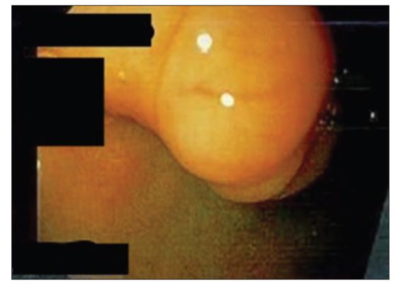
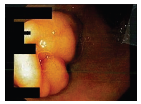
An EGD under general anesthesia was performed for possible endoscopic mucosal resection of the polypoid mass in the duodenum with possible open laparotomy if necessary. The lubricated endoscope was introduced, and the oropharynx was easily intubated with normal length. The Z line was noted at 38 cm. On entrance of the stomach, the scope was retroflexed and the cardia appeared normal with no evidence of a hiatal hernia. The scope was advanced into the antrum, which appeared normal, then the pylorus was intubated and the duodenum was traversed. The polypoid mass was well visualized in the first part of the duodenum appearing to be at least 4 cm [Figures 1 and 2]. On inspection, the remainder of the duodenum appeared to be normal. At this time, an endoscopic mucosal resection was performed. Two pink, polypoid, and soft rubbery excisions were retrieved using baskets that measured 1.8 cm X 1.4 cm X 1.2 cm and 2 cm X 1.4 cm X 0.9 cm. The base of the polypectomy site looked intact with no residual mass. The scope was retracted back into the stomach for a second look. The stomach was desufflated and the scope was removed with a second visualization of the mucosal surfaces on the way out. At this point, the procedure was deemed complete and the patient was awakened in stable condition for further recovery. The patient was placed on a proton-pump inhibitors BID and clear liquid diet and was advanced to normal diet on post-operative day 1 and was subsequently discharged.
Histopathology findings were consistent with chronic inflammation of the duodenal mucosa and an underlying polypoid mass of hyperplastic Brunner gland tissue, consistent with a Brunner's gland adenoma.
DISCUSSION
Brunner glands are normal submucosal cells that are most abundant in the proximal duodenum and can extend to the proximal jejunum [8]. These glands are made up of secretory units of epithelial tubules that secrete a protective and lubricating substance for the duodenum [9]. The mucous contains alkaline fluid, to protect the mucosa from gastric acidic secretions, a glycoprotein, that coats the intestinal mucosa, and enterogastrone, which inhibits gastric acid secretion [8]. It has been postulated that repeated duodenal mucosal injury either due to acidic gastric secretions or Helicobacter pylori infections are the cause of Brunner gland hyperplasia [10]. Brunner's adenoma is usually asymptomatic and found indecently during endoscopy. When the patient becomes symptomatic, it is usually due to the hamartoma becoming large enough to cause signs of obstruction and even hemorrhage [7]. Only 11% of patients with a case of Brunner's adenoma are asymptomatic [11]. For diagnosis, CT scans seem to be ineffective unless the mass is large enough to visualize. The mainstay of diagnosis and treatment, according to a study in Korea, is to perform and EGD polypectomy of Brunner's adenomas. Patients with large adenomas that have been unsuccessful with EGD or if the size is too large, must undergo open laparotomy for removal [12]. Whether to remove an asymptomatic hamartoma is not clear, their increasing size may result in complications of obstruction or hemorrhage as well as a potential for malignant transformation. This is reason enough for removal [13].
In conclusion, not only is EGD with endoscopic mucosal resection a generally safe and low-risk procedure, but studies have shown also excellent post-operative outcomes with no reoccurrences with both endoscopic mucosal resection and open laparotomy for removal [12]. While it remains controversial whether an asymptomatic Brunner's gland adenoma needs removed, removal of suspected Brunner adenomas is important to confirm diagnosis and prevent future complications such as obstruction, bleeding, or malignant transformation.
REFERENCES
- Michael JO, Kunitake H. Epidemiology, Clinical Features, and Types of Small Bowel Neoplasms. Available from:www.uptodate.com/contents/epidemiology-clinicalfeatures-and-types-of-small-bowel-neoplasms [Last accessed on 2018 Jan 05].
- Wang Y, Shi C, Lu Y, Poulin EJ, Franklin JL, Coffey RJ, et al. Loss of lrig1 leads to expansion of Brunner glands followed by duodenal adenomas with gastric metaplasia. Am J Pathol 2015;185:1123-34.
- Gotoh N. Feedback inhibitors of the epidermal growth factor receptor signaling pathways. Int J Biochem Cell Biol 2009;41:511-5.
- Hanahan D, Weinberg RA. Hallmarks of cancer: The next generation. Cell 2011;144:646-74.
- Wang Y, Poulin EJ, Coffey RJ. LRIG1 is a triple threat: ERBB negative regulator, intestinal stem cell marker and tumour suppressor. Br J Cancer 2013;108:1765-70.
- Stewart ZA, Hruban RH, Fishman EF, Wolfgang CL. Surgical management of giant brunner's gland hamartoma: Case report and literature review. World J Surg Oncol 2009;7:68.
- Rocco A, Borriello P, Compare D, De Colibus P, Pica L, Iacono A, et al. Large Brunner's gland adenoma: Case report and literature review. World J Gastroenterol 2006;12:1966-8.
- Carrie W, Matin A, Syed MA, Levi GS, Carr-Locke DL. When Brunner glands are not so innocent. Medscape Medscape Gastroenterol 2012. Available from: http://www.medscape.com/viewarticle/775988_1. [Last accessed on 2018 June, 08].
- Krause WJ. Brunner's glands: A structural, histochemical and pathological profile. Prog Histochem Cytochem2000;35:259-367.
- Kovacevic I, Ljubicic N, Cupic H, Doko M, Zovak M, Troskot B, et al. Helicobacter pylori infection in patients with Brunner's gland adenoma. Acta Med Croatica 2001;55:157-60.
- Walden DT, Marcon NE. Endoscopic injection and polypectomy for bleeding Brunner's gland hamartoma: Case report and expanded literature review. Gastrointest Endosc 1998;47:403-7.
- Gao YP, Zhu JS, Zheng WJ. Brunner's gland adenoma of duodenum: A case report and literature review. World J Gastroenterol 2004;10:2616-7.
- Park JH, Park CH, Park JH, Lee SJ, Lee WS, Joo YE, et al. The safety and usefulness of endoscopic polypectomy for treatment of Brunner's gland adenomas. Korean J Gastroenterol 2004;43:299-303.