Asclepius Medical Case Reports
Volume 1, Issue 1, 2018, Page No: 1-2
Mamelon Paget Disease about a Case
Houda Moustaide, Hanane Saadi, Hafsa Taheri, Saad Benkirane, Ahmed Mimouni
Department of Obstetric and Gynecology, University Hospital Mohammed VI, Oujda, Morocco.
Citation : Moustaide H, Saadi H, Taheri H, Benkirane S, Mimouni A. Mamelon Paget Disease about a Case. Asclepius Med Case Rep 2018;1(1):1-2.
Abstract
Tumors of the nipple-areolar plate are rare. Paget's disease is one of them; it corresponds to the invasion of the nipple epidermis by carcinoma in situ. It represents 1-3% of mammary tumors isolated or associated with underlying breast cancer in 80% of cases. Its diagnosis is histological, and it is done by the cytological scraping of the nipple.
Disease, Paget, rare, tumor
INTRODUCTION
Paget's disease is a malignant skin disorder affecting the nipple and areola, almost always associated with breast carcinoma. It is clinically manifested by fairly welldefined erythematous plaques, sometimes already associated with a palpable mammary tumor. The biopsy is necessary for the positive diagnosis. The treatment is surgical and depends on the nature and extent of the underlying tumor.
The aim of our work is to determine through a case of nipple Paget's disease collected in our training, and a review of the literature, the epidemiological, diagnostic, therapeutic, and prognostic factors of nipple Paget's disease.
CASE REPORT
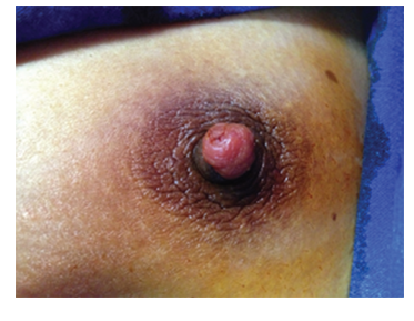
This is a 35-year-old female patient with two live children with no notable pathological history who has had a reddish lesion of the nipple and pruriginous of the left nipple for the past 4 months, has a tumefaction of the eczematous 1 cm erythematous left nipple, with no palpable mammary nodule or nipple discharge, the ganglionic areas are free [Figure 1]. Breast ultrasound and bilateral mammography were normal. The nipple biopsy showed a fairly well-differentiated Paget's disease of the left nipple with no invasiveness.
The treatment consisted of a lumpectomy with the left areola-nipple plate whose anatomopathological examination revealed nipple Paget's disease associated with an intraductal carcinoma of intermediate grade interesting the left breast with healthy resection areas without homes without microinfiltration. The patient received 33 radiotherapy sessions postoperatively.
Figure 1: Photographic image showing 1 cm red mamelonary lesion without nodule under jacent
DISCUSSION
Paget's disease is a rare and unknown pathology. It represents only 0.7-4.3% of all breast cancers, and its incidence has been decreasing for several years. It affects women around the age of 60. Its development is insidious, unilateral, and centrifugal. Its macroscopic appearance is that of exudative and crustal eczema of pink to red color. When it is usually associated with breast carcinoma, it is palpable only in 33% of cases at the time of diagnosis [1].
Clinically, it often poses a differential diagnosis problem with irritative dermatitis, eczema, scabies, and psoriasis. However, some stigmas favor these pathologies, in particular, their bilaterality, the presence of other localizations, their evolution by outbreaks and their pruriginous nature. A radiodermatitis can also simulate a Paget's disease, but the antecedents of irradiation make it possible to easily evoke this diagnosis, as well as other malignant pathologies such as basal cell carcinoma pagetoid or melanoma [1,2].
The anatomopathological diagnosis is made by cutaneous biopsies, rarely on the mastectomy specimen, by standard histology and immunohistochemistry.
The lesions are visible on mammography in 35-78% of cases. The most frequent anomaly is the presence of microcalcifications suggestive of Paget when they are localized in intra- or basi-nipple, punctiform or linear in ductus disposition (12% of microcalcifications), or even grouped in foci or beaches. Paget's nipple disease is associated with a low sensitivity of clinical examination and mammography/ultrasound for the detection of breast carcinoma associated. Several studies have shown that in cases of nipple Paget's disease without palpable mass and without mammographic abnormality, histology accounted for approximately 43% of occult cancers, 33-75% of noninvasive cancers, and 5 to 9% of invasive cancers. In a recent study, the sensitivity of mammography was only 34% of cases of surgically confirmed cancers. In another series, the sensitivity of the mammogram was better, but the provision of ultrasound to the mammogram was very relative, increasing the undetected cancer rate from 15% to 13%. Pre-operative breast magnetic resonance imaging is useful if conservative surgery is considered because of a high rate of occult cancer in mammography and ultrasound [2].
Mastectomy has long been regarded as the gold standard treatment for Paget's disease given the potential multifocality.
Some teams have proposed conservative surgical treatment, penectomy, associated with radiotherapy of the entire mammary gland in selected patients, with no palpable mass or mammographic abnormality. Studies have shown that longterm survival is similar in these patients compared to those who have had a mastectomy. However, this conservative attitude is also controversial given the underestimation of mammography for the detection of carcinomas and their multifocal or multicentric character [3,4].
The prognosis is influenced by the presence of underlying breast cancer and its invasiveness or non-invasiveness. Survival at age 15 is 88% for patients without underlying neoplasia, 61% for patients with infiltrating ductal carcinoma, and 94% with cancer in situ. Paget's disease associated breast cancers have a poorer prognosis than "classic" breast cancers. They are usually high grade, large (>2 cm) and lymph node disseminated [4].
CONCLUSION
Persistent unilateral nipple necrosis should be suspected of nipple Paget's disease. Its diagnosis is done by a simple cytological scraping of the nipple, its treatment is surgical and depends on the underlying lesions, and the prognosis is influenced by the invasiveness or not of an underlying mammary cancer [3].
References
- Geffroy D, Doutriaux-Dumoulins I, Labbe-Devilliers C, Meingan P, Houdebine S. Maladie de paget du mamelon et principaux diagnostics differentiels. J Radiol 2011;92:889-98.
- Nicholson BT, Harvey JA, Cohen MA. Nipple-areolar complex: Normal anatomy and benign and malignant processes. Radiographics 2009;29:509-23.
- Kinoshita S, Yoshimoto K, Kyoda S, Hirano A, Shioya H, Kobayashi S, et al. Malignant melanoma originating on the female nipple: A case report. Breast Cancer 2007;14:105-7.
- Dalberg K, Hellborg H, Warnberg F. Paget's disease of the nipple in a population based cohort. Breast Cancer Res Treat 2008;111:313-9.