Melanoma of the Palate: A Case Report and Literature Review
Allouane Mohamed Amine, Lekhbal Adil, Laababsi Rabii, Rouadi Sami, Abada Reda Lah, Roubal Mohamed, Mohamed Mahtar
Citation : Amine AM, Adil L, Rabii L, Sami R, Lah AR, Mohamed R, Mahtar M. Melanoma of the Palate: A Case Report and Literature Review. Asclepius Med Case Rep 2018;1(1):1-3.
Mucous melanoma accounts for 1% of melanomas. The palate and the labial mucosa are the most affected. It is a dark prognostic
tumor, the treatment associated with surgery and radiotherapy in the localized stages. We report the case of a palate melanoma in a 47-year-old patient without distant metastases.
Mucosal melanoma, oral cavity, palate, prognosis
INTRODUCTION
Cervicofacial mucosal melanoma accounts for 55% of all mucosal melanomas [1]. In the oral cavity, the palate and alveolar gingiva are the most common sites, followed by the labial mucosa, oral mucosa, and tongue[2].
Mean age at diagnosis is between 65 and 70 years later than in cutaneous melanomas [3,4]. They are often detected late, which aggravates their prognosis.
CASE REPORT
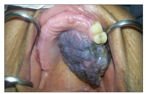
This is a 47-year-old woman with no particular pathological history who consulted for an endobuccal mass that appeared 5 months ago. The exobuccal examination shows asymmetry of the face, and the endobuccal examination reveals a budding tumor, blackish in color, bleeding on contact, sitting on the level of the hard palate, and exceeding the median line, which reaches the soft palate and the upper dental arch [Figure 1]. The cervical examination does not find lymphadenopathy.
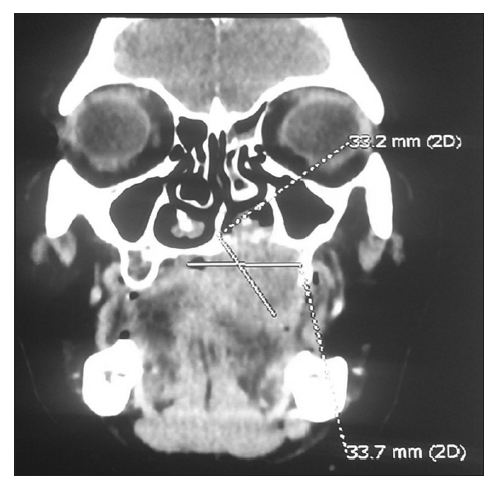
The computed tomography (CT) scan of the face showed a large mass at the hard palate with significant osteolysis and which reaches the ipsilateral nasal cavity [Figure 2].
The histopathological examination confirms the diagnosis of melanoma. The extension assessment did not reveal the existence of metastases.
The patient underwent surgical treatment (maxillectomy with lymph node dissection) associated with radiotherapy, and an obturator prosthesis was performed.
DISCUSSION
Mucous melanomas of the oral cavity are rare. They are most often found in the hard palate, the soft palate, and the upper gum [1].
Macroscopically, it is most often a pigmented lesion, in 30% of cases, the pigmentation would precede tumor onset [5,6]. Bleeding, pain, tooth mobility, or delayed healing after extraction may occur.
Extension assessment includes cervicofacial CT or magnetic resonance imaging, thoracoabdominal CT scan, and bone scintigraphy.
The etiology of melanoma of the oral mucosa is not yet known. Risk factors could be tobacco and trauma [4,6].
In 25% of cases, there is cervical lymphadenopathy, distant metastases are mainly pulmonary, mediastinal, cerebral,hepatic, and bone [4].
The most commonly used classification for mucosal melanoma is Ballantyne, which distinguishes three stages, Stage I corresponds to limited involvement in the primary lesion, Stage II has nodal invasion, and Stage III has distant metastatic disease [7]. Prasad subdivided Stage I of Ballantyne in three levels, the first corresponding to melanomas in situ, the second to lamina propria involvement, and the third to deep tissue invasion (muscle, bone, or cartilage) [8].
The treatment is not consensual. Wide excision, whether or not associated with lymph node dissection, is often recommended. Although these tumors are resistant to radiotherapy, radiotherapy is proposed in cases of nodal invasion with capsular rupture.
The indication for cervical dissection depends on the presence of lymphadenopathy and primary localization. More than 25% of patients with melanoma of the oral cavity have lymph node metastases, a systematic dissection is justified and all the more so that the nodal recurrence rate is high (70%)[9]. The first-line radiotherapy may be offered for unresectable melanoma or for inoperable patients.
The prognosis is favorable when the diagnosis is early and the treatment is adequate. However, many patients have an advanced stage of diagnosis, and the rate of distant metastases remains high even for patients diagnosed at an early stage, and the 5-year survival rate ranges from 5 to 20% [4,5].
CONCLUSION
Cervicofacial mucosal melanomas are rare and have a poor prognosis. The diagnosis can sometimes be difficult [10]. The treatment of localized stages is radiosurgical. For advanced stages, management remains palliative. However, a biopsy-excision with margins of excision of any pigmented lesion of the oral cavity is essential, especially in the palate.
REFERENCES
- Chang AE, Karnell LH, Menck HR. The American college of surgeons' commission on cancer and the American cancer society. The national cancer data base report on cutaneous and noncutaneous melanoma: A summary of 84,836 cases from the past decade. Cancer 1998;83:1664-78.
- Troussier I, Baglin AC, Marcy PY, Even C, Moya-Plana A, Krengli M, et al. Melanomes muqueux de la tete et du cou: Etat actuel des pratiques et controverses. Bull Cancer 2015;102:559-67.
- Gru AA, Becker N, Dehner LP, Pfeifer JD. Mucosal melanoma: Correlation of clinic pathologic, prognostic, and molecular features. Melanoma Res 2014;24:360-70.
- Hicks MJ, Flaitz CM. Oral mucosal melanoma: Epidemiology and pathobiology. Oral Oncol 2000;36:152-69.
- Meleti M, Leemans CR, Mooi WJ, van der Waal I. Oral malignant melanoma: The amsterdam experience. J Oral Maxillofac Surg 2007;65:2181-6.
- Rapini RP, Golitz LE, Greer RO Jr., Krekorian EA, Poulson T. Primary malignant melanoma of the oral cavity. A review of 177 cases. Cancer 1985;55:1543-51.
- Michel J, Perret-Court A, Fakhry N, Braustein D, Monestier S, Richard MA, et al. Sinonasal mucosal melanomas: The prognostic value of tumor classifications. Head Neck 2014;36:311-6.
- Prasad ML, Patel SG, Huvos AG, Shah JP, Busam KJ. Primary mucosal melanoma of the head and neck: A proposal for microstaging localized. Stage I (lymph node-negative) tumors. Cancer 2004;100:1657-64.
- Patrick RJ, Fenske NA, Messina JL. Primary mucosal melanoma. J Am Acad Dermatol 2007;56:828-34.
- Baujat B, Thariat J, Baglin AC, Costes V, Testelin S, Reyt E, et al. Rare tumors of the head and neck; On behalf of the REFCOR, the French network of rare head and neck tumors. Bull Cancer 2014;101:411-23.