Unusual Repetitive Vasculitis Derived Splinter Hemorrhage Case in the One Finger
Suna Aydin
Citation : Aydin S. Unusual Repetitive Vasculitis Derived Splinter Hemorrhage Case in the One Finger. Asclepius Med Case Rep 2018;1(1):1-3.
Vasculitis is an inflammatory process that affects the vessel wall and leads to the narrowing of the lumen due to thickening or to total occlusion due to thrombosis resulting in tissue ischemia and necrosis. Here, it was reported the case of a 55-year-old man with vasculitis derived splinter hemorrhage in the finger and was initially treated with acetylsalicylic acid and acetylsalicylic acid allergy was developed within 10 min. Later on, clopidogrel 75 mg was given to prevent the vasculitis derived splinter hemorrhage, which developed in the third fingernail bed, to obstruct the capillaries. Right diagnosis and treatment of vasculitis on time are important since a delay in diagnosis may impact the outcome significantly including thrombosis and vascular occlusion, which might be resulted in the mortality and morbidity.
fingernail bed, splinter hemorrhage, thrombosis, vasculitis.
INTRODUCTION
Vasculitis is known as inflammation of blood vessels [1]. There have been known different variable forms in world [1-4]. Some bladder and kidney cancers have been reported to be associated with vasculitis [5]. There might be also linked with splinter hemorrhage. Various biomarkers are used in detection [6].
CASE REPORT
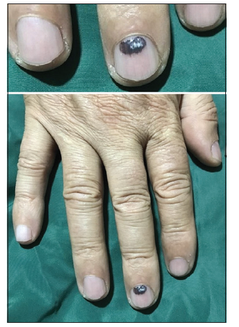
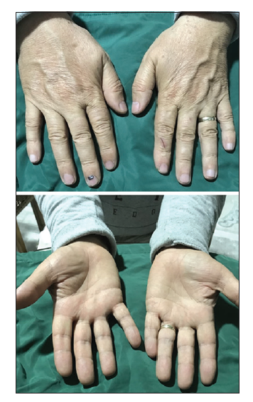
A 55-year-old male patient admitted to our clinic with hemorrhage spontaneously developing in the right-hand third phalanx (finger) nail bed due to cold exposure [Figure 1]. The patient stated that this hemorrhage was in a repetitive manner for about 35 years. The diagnosis of his referral clinical manifestation was Raynaud's syndrome. In his medical history, it was recorded that the patient was admitted to the emergency service for the cause of severe vertigo due to cold exposure 14 years ago. It was also seen in his anamnesis that following the pain associated with the cold sensation in the sacral region; hemorrhage-like color change was occurred at his third phalanx nail bed. The patient also said hemorrhage in his finger appeared within 1 week following the pain felt in the sacral region. When the third phalanx of right and left hands was compared, the third phalanx of the right hand was found to have a bigger diameter [Figure 2]. This diameter difference was detected in a clinic in the USA for the 1st time 26 years ago. The right-hand third finger biopsy was taken and no pathological findings were found. The patient also said that he had applied a numerous clinic until so far and diagnosed "Raynaud's syndrome." His last visit our hospital (emergency center) was due to dizziness, nausea, and vomiting and then consulted cardiovascular department to treat him. Then, acetylsalicylic acid was administered to the patient for antiaggregant therapy,[7] and acetylsalicylic acid allergy was developed within 10 min, and emergency intervention was performed due to angioneurotic edema. Later on, clopidogrel 75 mg was given to prevent the hemorrhage, which developed in the third fingernail bed, to obstruct the capillaries.
DISCUSSION
When we put all of the above anamnesis data together and evaluate it, although Raynaud's syndrome was evaluated in the first place[7] when the patient's finger structures were examined, presence of no loss of pain and sensory perception ability, presence of no response to the cold water stress test, and absence of color change in the nail as a result of cold and emotional stress exposure excluded the diagnosis of Raynaud syndrome [Figure 1].[8] Therefore, the case suggests being splinter hemorrhage[9] rather than Raynaud syndrome [8] Because the presence of dark red-brown linear bleeding in the nail can be seen in splinter hemorrhage and no physical trauma story was observed in this case. In such cases, the development of hemorrhage in the right third phalanx nail bed with recurrent intervals due to coldness other than dizziness and vomiting did not lead to a vital condition. However, initiation of clopidogrel by referral to the clinic at the first sensation of pain in the sacral region, to remove any potentially damaging effects of such cases, will protect the patient from potentially life-threatening effects. Up to this time, pain in the sacral area due to cold was only found in the right-hand third phalanx.
It is also understood that the appearance of this hemorrhage within 1 week following the pain felt in the sacral region is directly related to the growth rate of the nail. Because the nail growing is about 3.47 mm (0.11 mm/day) in 1 month [10]. The determination of hemorrhage in the nail bed within 1 week following exposure to the cold confirms this grow within 1 week. The occurrence of dizziness due to the abovementioned coldness suggests that the vasculitis derived splinter hemorrhage [8] in this nail is associated with dizziness, nausea, and vomiting.
Based on all this information, it is understood that the development of hemorrhage in the nail is not a lifethreatening condition. However, against all possibilities (including capillary occlusion), we think that giving the above treatment is important for patients. An other words, this repetitive vasculitis derived splinter hemorrhage is rare and heterogeneous disease that appears all the time with hemorrhage in the right third phalanx nail bed with recurrent intervals depending on the cold feeling in the sacral region and pain in the kidney. Recognition of this case by clinicians is important since a delay in diagnosis may impact the outcome significantly (such as thrombosis and vascular occlusion), which might be resulted in the mortality and morbidity. Thus, an overall clinical assessment is important in the initial evaluation of patients with suspected vasculitis derived splinter hemorrhage. Furthermore, differential diagnosis with secondary causes of vasculitis (e.g. bacterial infection) and with vasculitis mimics should be taken into account in a patient with suspected vasculitis.
CONCLUSION
Although the differential diagnosis is broad, prostate origin must be entertained in an elderly male presenting with supraclavicular lymphadenopathy. Metastatic cancers of the neck are more commonly from a squamous cell origin. In this case, we presented a patient with progressively enlarging the left supraclavicular lymphadenopathy. The importance of immunohistochemical staining with PSA cannot be understated.
REFERENCES
- Sagheb K, Menz A, Albrich S, Taylor K, Hess G, Walter C. Supraclavicular metastases from distant primary solid tumors: A retrospective study of 41 years. J Maxillofac Oral Surg 2017;16:152-7.
- Park E, Stroie F, McArdle B, Psutka S. Metastatic adenocarcinoma of prostate presenting as supraclavicular and bulky generalized lymphadenopathy with a benign digital rectal exam. Urol Case Rep 2017;13:128-30.
- Arumugam S, Kanakasabapathy S, Farman MJ. Combined androgen blockade responsive prostate carcinoma with extensive nodal metastases with Virchow's node. J Evol Med 2017;6:661-4.
- Lin YY, Lin DS, Kang BH, Lin YS. Neck mass as the first presentation of metastatic prostatic adenocarcinoma. J Chin Med Assoc 2011;74:570-73.
- Wang HJ, Chiang P, Peng J, Yu T. Presentation of prostate carcinoma with cervical lymphadenopathy: Report of 3 cases. Chang Gung Med J 2004;27:840-4.
- Jones H, Anthony PP. Metastatic prostatic carcinoma presenting as left sided cervical lymphadenopathy: A series of 11 cases. Histopathology 1992;21:149-54.
- Davarci M, Gokce A, Guven ED, Yalcinkaya F, Esen H, Sevine A. Metastatic prostate adenocarcinoma presenting with supraclavicular lymphadenopathy: A report of two cases. Contemp Oncol 2012;16:53-5.
- Pinaquy J, Allard J, Cornelis F, Pasticier G, De Clermont H. Unusual lymph node metastasis of prostate cancer detected with 18F-flourocholine PET/CT. Clin Nucl Med 2015;40:e255-57.
- American Cancer Society. About Prostate Cancer 2017. Atlanta, GA: American Cancer Society; 2017.
- Siegel R, Miller K, Jemal A. Cancer Statistics 2017. Cancer 2017;67:7-30.
- Steele CB, LI J, Hueng B, Weir HG. Findings from CONCORD-2 study. Cancer 2017;123:5160-77.
- Moyer VA. On behalf of the U.S. preventive services task force. screening for prostate cancer: U.S. preventive services task force recommendation statement. Ann Intern Med 2012;157:120-34.
- Bubendorf L, Schopfer A, Wagner V, Sautar G, Moch H, Gaser TC. Metastatic patterns of prostate cancer: An autopsy study of 1589 patients. Human Pathol 2000;31:578-83.
- Hematpour K, Bennett CJ, Rogers D, Head CS. Supraclavicular lymph node: Incidence of unsuspected metastatic prostate cancer. Eur Arch Otorhinolaryngol 2006;263:572-4.
- Balikci HH, Gurdal MM, Ozkul MH, Uvacin O, Kara N. Neck masses: Diagnostic analysis of 630 cases in Turkish population. Eur Arch Otorhinolaryngol 2013;270:2953-58.