Did Stem Cells Play Any Role in the Curious Case of Transplantation of Cancer in 1874?
Wilson I. B. Onuigbo
Citation : Onuigbo WIB. Did Stem Cells Play Any Role in the Curious Case of Transplantation of Cancer in 1874?. Asclepius Med Res Rev 2018;1(1):1-2.
It was stated recently that the concept of cancer stem cells "has directed scientific communities toward a different wide new area of research field," and that "The cancer stem cells may be able to answer some of the questions related to a cancer growth." Accordingly, this paper recollects the odd presentation of cancer in 1874 and wonders if the stem cell system was at work then.
Cancer, case report, history, metastasis, stem cells
INTRODUCTION
In their thought-provoking review on the role of stem cells in cancer,[1] Gagar's associates mentioned not only that they are "now considered as backbone in the development of the cancer; and their role in carcinogenesis" but also that they "may be able to answer some of the questions related to a cancer growth."
Accordingly, the historical approach is offered here for consideration on account of the sheer diversity of the growths although two primaries were probably involved [2].
HISTORICAL TEXT
James Goodhart, in 1874, reported to the Pathological Society of London the case of EJ, a woman whose ovary had been removed a month earlier. At autopsy, the findings were variegated. Incidentally, the word "tubercle" was used, this being, as I reported elsewhere,[3] coterminous with cancer and tuberculosis!.
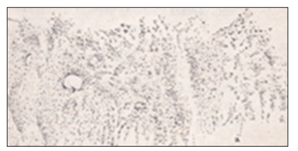
Both pleura and lungs were affected. They were most thickly set toward the bases. At microscopy, the growth was of "small roundish nuclei, not unlike an interstitial pneumonia." The ileum had "two or three small cancer tubercles under the peritoneum." The liver contained in its substance a few secondary "cancer nodules." The peritoneum near to the pedicle of the diseased ovary was studded. The pedicle itself was a mass of new growth. The caliber of part of the tube was dilated, and "full of cream-like juice."
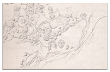
Uterus large. On the posterior wall of its cavity was a fleshy looking mass, sessile, but projecting from the surface. It had a soft vascular appearance and yielded copious juice on section. It was apparently a thickening of the mucous coat. The mucous membrane elsewhere was soft, and undergoing a cancerous change; uterine muscle looked healthy. Microscopically, however, this was not so [PI. VIII, Figure 2]. The muscular tissue is extensively infiltrated by a cellular growth. The mucous coat is much thickened, has entirely lost all appearances of columnar epithelium, and is replenished by a free growth of epithelium-like cells, similar to those of the growth in the ovary.
The lumbar lymph nodes were cancerous. One kidney had a cancerous nodule in it. The ovary was large and contained one or two small cancer nodules on it, and also in its substance.
The interest of the case lay in the facts:
- First, that it was a good specimen of autoinoculation of cancer, parallel to a case brought before the Society by Dr. Moxon, and published in a late volume of the "Transactions," where cancer about the larynx had been carried down the bronchi and had grown there.
- Second, it was a good specimen of the power cells inabnormal irritative states have on presumably healthy cells. Cells of bad habit from the ovary traverse the fallopian tube and so affect the nutrition or life action of the cells of the uterine mucous membrane that they throw off all allegiance to their former type, grow ovary tumor cellshenceforward, and not uterine mucous membrane cells.
- Third, this so-called spermatic influence of one cell on another was lost or not manifested in the lung. Thus, in it, the growth was more like an interstitial pneumonia than resembling the original ovarian tumor.
DISCUSSION
As the above details have shown, that medical master included figures whose cellular pictures were of two types of cancer cells. Therefore, I am persuaded that one of the primaries was pulmonary going by the statement made in the third interesting type above.
It is to be noted that Willis,[4] as is his practice, mentioned this particular historical article as follows:
The occurrence of surface implantation in the uterine endometrium is difficult to establish; but from my study of recorded cases, I believe that genuine implant metastases from ovarian and tubal tumors probably do occur in the endometrium, for examples, discussion and literature consult Goodhart.
CONCLUSION
It is salutary to mention that, when consulting the medical masters of yester years, it is well to be reminded not only that surgical oncology was advancing during their time[5] but also that acknowledgments of previous writers were occurring [6]. Thus, the old citation was from Moxon's report culled from a volume of the transactions. In this context, the problem of resolving any doubts still continues. For instance, concerning the present ovary lung axis, Young and Scully handled such problems with regard to the interpretation of ovarian metastases and cancer of the lung [7].
REFERENCES
- Corrado D, Thiene G, Nava A, Rossi L, Pennelli N. Sudden death in young competitive athletes: Clinicopathologic correlations in 22 cases. Am J Med 1990;89:588-96.
- Basso C, Corrado D, Marcus FI, Nava A, Thiene G. Arrhythmogenic right ventricular cardiomyopathy. Lancet 2009;373:1289-300.
- Murray B. Arrhythmogenic right ventricular dysplasia/ cardiomyopathy (ARVD/C): A review of molecular and clinical literature. J Genet Couns 2012;21:494-504.
- Basso C, Corrado D, Thiene G. Cardiovascular causes of sudden death in young individuals including athletes. Cardiol Rev 1999;7:127-35.
- Valente M, Calabrese F, Angelini A, Basso C, Nava A, Rossi L, et al. In vivo evidence of apoptosis in arrhythmogenic right ventricular cardiomyopathy. Am J Pathol 1998;152:479-84.
- Thiene G, Nava A, Corrado D, Rossi L, Pennelli N. Right ventricular cardiomyopathy and sudden death in young people. N Engl J Med 1988;318:129-33.
- Marcus FI, Fontaine GH, Guiraudon G, Frank R, Laurenceau JL, Malergue C, et al. Right ventricular dysplasia: A report of 24 adult cases. Circulation 1982;65:384-98.
- Pinamonti B, Sinagra G, Salvi A, Di Lenarda A, Morgera T, Silvestri F, et al. Left ventricular involvement in right ventricular dysplasia. Am Heart J 1992;123:711-24.
- Foale RA, Nihoyannopoulos P, McKenna WJ, Oakley CM, Krikler DM, Rowland E, et al. Right ventricular abnormalities in ventricular tachycardia of right ventricular origin: Relation to electrophysiological abnormalities. Br Heart J 1986;56:45-54.
- Jain R, Dalal D, Daly A, Tichnell C, James C, Evenson A, et al. Electrocardiographic features of arrhythmogenic right ventricular dysplasia. Circulation 2009;120:477-87.
- Bayar N, Arslan S, Koklu E, Cagirci G, Cay S, Erkal Z, et al. The importance of ECG findings in the diagnosis of atrial septal defect. Kardiol Pol 2015;73:331-6.
- Rojas CA, El-Sherief A, Medina H. Embryology and developmental defects of the interatrial septum. Am J Roentgenol 2010;195:1100-4.