Effect of Riboflavin on Push-out Bond Strength between Fiber Post and Root Dentin using Adhesive Cement - An in vitro Study
Asma Altaf1, Lekha Santhosh1, A. Srirekha1, Srinivas Panchajanya1, T. Jaykumar1
Citation : Altaf A, Santhosh L, Srirekha A, Panchajanya S, Jaykumar T. Effect of Riboflavin on Push-out Bond Strength between Fiber Post and Root Dentin using Adhesive Cement - An in vitro Study. J Clin Res Dent 2019;2(1):1-5.
Durability of the bond between the fiber post and the root dentin are an important issue for providing long-term clinical success. The aim of this study was to assess the influence of riboflavin (RF) application on bond strength of fiber posts to root canal dentin using a self-etch adhesive luting system.
A total of 40 single-rooted teeth freshly extracted for the orthodontic purpose were decoronated below the cementoenamel junction, and standardized to 14 mm in length. Post space preparation was done up to No. 3 Peeso reamer. Teeth were randomly divided into two Groups: Group 1: Pre-treatment with etchant and RF (n = 20) and Group 2: No pre-treatment with RF (n = 20). Both the Groups were restored with fiber posts bonded with a self-etch adhesive luting cement. Teeth were then stored in distilled water for a period of 3 months at 37°C. 1 mm slices of the coronal and middle third of root were obtained, and push-out bond strength testing was done with a universal testing machine. Failure patterns were assessed under a stereomicroscope at *20 magnification.
The values were tabulated, and statistical analysis was done with the Chi-square test and independent t-test. There was a statistically significant difference in push-out bond strength between the two groups (P < 0.001) and also between the coronal and middle third region.
Within the limitations of the study, it can be concluded, that pre-treatment with RF during bonding procedure preserves the bond strength of fiber posts to root dentin.
Collagen cross-linking, glass fiber post, matrix metalloproteinase's inhibitor, push-out bond strength riboflavin, self-etch adhesive system,Dentistry
INTRODUCTION
Endodontically treated teeth often loose substantial tooth structure from previous caries, pre-existing restorations, and/or endodontic treatment and hence the need of an additional post and core as a post-endodontic restoration arises for the retention of the crown. A non-metallic fiber post is a better choice than metal as it improves stress distribution and decreases chances of fracture, under function.
The durability of the bond between the fiber post and the root dentin is an important issue for providing long-term clinical success. The hydrolytic degeneration of resin and degradation of collagen leads to bond failure. The structural integrity and mechanical properties of collagen fibrils directly affect the quality of bond strength and its durability. Thus, to strengthen collagen fibrils at resin-dentin interface, collagen crosslinking agents have been used. Exposed collagen after etching procedure is gradually degraded by matrix metalloproteinase's (MMP) and hence there is also a role of MMP inhibitors in strengthening the dentin adhesive bond[1].
Riboflavin (RF) is one such compound that has ability to produce free radicals when photoactivated and it is biocompatible. Several corneal (eyes) studies, both in vitro and in vivo with RF, have shown effective results by strengthening the collagen[2-4]. Studies have shown that RF, when applied during the bonding procedure in coronal dentin, increases the resin-dentin bond strength and decreases the degradation of collagen breakdown by reducing the destructive potential of MMPs and by its cross-linking ability[5-8]. Till date, very few studies have been done on effect of RF on root dentin[9,10].
Hence, the aim of this in vitro study was to assess the influence of photoactivated 1% RF application during the bonding procedure on push-out bond strength of glass fiber posts to root canal dentin using adhesive luting system after storage of the specimens for a period of 3 months. The null hypothesis states that there will be no difference in the push-out bond strength of fiber post to root dentin using an adhesive cement with or without the application of photoactivated RF during the bonding procedure.
MATERIALS AND METHODS
Single-rooted premolars (extracted due to orthodontic reason) with fully developed mature apices with straight canals and round cross-section were selected and stored in 0.1% thymol solution. Ethical clearance was obtained from the Ethical Committee of the institutionbefore the study. Teeth with morphological defects, previous endodontic treatment, cracks or fracture lines, or calcified canals were excluded. Teeth kept for not more than 3 months after debridement were taken for the study and were washed in running water to eliminate thymol residues before initiating the study. The sample size of 20 for each group was estimated using the GPower software v. 3.1.9.2 for power of the study at 80%.
Each tooth was decoronated below the cementoenamel junction perpendicular to the longitudinal axis using a slow-speed, water-cooled diamond disk such that they were cut to a uniform length of 14 mm from the apical end. Pulp tissue was removed using files #10K and #15K file (Mani. Inc., Japan). Proper cleaning and shaping were done with Pro Plus Gold rotary files up to 30.6% using X-mart endomotor with speed of 350 rpm and torque 2.2 N cm. Normal saline, 3% sodium hypochlorite and 17% ethylenediaminetetraacetic acid (EDTA) were used as irritants. The apices of the teeth were sealed with reinforced zinc oxide eugenol (IRM, DENTSPLY Caulk), a provisional filling material.
Peeso reamers (Mani. Inc., Japan) sizes 1, 2, and 3 were used to prepare the post space of 10 mm length into which a fiber post was to be fitted. The canals were irrigated during post space preparation followed by a final irrigation protocol of 5 ml of 3% sodium hypochlorite (Vensons India), 17% EDTA solution (pulp dent EDTA solution, pulp dent corporation), and 0.9% normal saline. The fit of the glass fiber post of diameter 1.2 mm (Roca AAA fiber post,) was then checked inside the root canal. The canal preparations were etched with 37% phosphoric acid with an applicator tip for 15 s followed by rinsing and drying with paper points.
The teeth were randomly divided into two Groups:
Group 1: Root canals treated with 1% RF and restored with glass post of diameter 1.2 mm (n = 20)
Group 2: Non-RF treated root canals restored with the glass fiber post (n = 20).
About 1 g RF (zenith nutrition VB7115) in powder form (yellow powder) was mixed in 100 ml of distilled water solution to obtain the 1% RF solution (the RF solution has to be prepared just before the application as it loses its property and becomes inactive within 4 h).
Group1: In this group, the custom prepared 1% RF was applied to the canal with the help of 27-gauge open-ended needle (care taken to avoid air bubble), light transmitting fiber post was placed in the canal and then photoactivated with blue light for 2 min with intensity of 600 mW/cm2 using conventional light curing unit. Fiber post was removed from the canal and excess was blot dried using paper points.
Subsequently, the fiber post was covered with the dual-cure resin cement (RelyXU2003MESPE), and then the post was seated inside the root canal with to and fro motion and then kept under the finger pressure for 20 s with excess cement removed. The dual-cure resin cement was polymerized for 40 s using the above-mentioned light curing unit.
Group 2: The teeth in this group were restored with fiber post in the similar way as in the Group 1 except for the pretreatment with RF.
A specimen of both the group was stored in distilled water for a period of 3 months, and after every 10 days, the distilled water was changed. Each root was mounted on acrylic block customized for the rotary microtome (Physilab, Germany) and then cut horizontally into 1 mm sections under wet condition. Sections were obtained from cervical third and middle thirds of the root. Each section was marked on its coronal side with an indelible marker. The thickness of each slice was reassessed using a digital caliper.
To evaluate the bond strength, a thin slice push-out strength test was performed with a universal testing machine (Multitest-i, Mecmesin, England). A load was applied in apicocoronal direction to the post surface that resulted in shear stresses along the luted interfaces. The diameter of the plunger was 1 mm to ensure that it contacted only the post during loading. Loading was performed at a crosshead speed of 1 mm/min until failure. The load needed to dislodge the posts was recorded in Newton (N). To express the bond strength in Mega Pascal's (MPa), the load at failure recorded in Newton's (N) was divided by the area (mm2) of the post/ dentin interface. The failure patterns were assessed under *20 magnifications with a stereomicroscope (ZOOMAR, Lawrence, and Mayo) and scores were given[11].
RESULTS AND STATISTICAL ANALYSIS
The force in Newton and push-out bond strength in MPa for both the groups was tabulated in Microsoft Excel. Statistical Package for the Social Sciences for Windows Version 22.0 Released 2013 Armonk, NY: IBM Corp. was used to perform statistical analyses. The level of significance was set at P < 0.05.
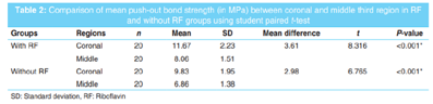
A statistically significant difference in push-out bond strength was found between the two groups [Table 1] and also between the coronal and middle third region in both the groups [Table 2].

Chi-square test was used to compare the failure scores between the two groups at the coronal and middle 3rd region. McNemar's test was used to compare the failure scores between a coronal and middle 3rd region in each group. However, no significant difference was found.
DISCUSSION
Badly broken down teeth are further weakened by the endodontic procedures designed to provide optimal access and by the restorative procedures required to rebuild the tooth. Loss of inherent dentinal fluid may also effect an alteration in tooth properties. Ensuring optimal anchorage while maintaining adequate root strength for the particular clinical situation can be challenging and the problems encountered have resulted in the development of different materials and techniques.
Fiber post has a modulus of elasticity similar to dentin, which allows them to flex with the root when under stress. This is believed to distribute the stresses more evenly throughout the tooth than metal posts, making the root less susceptible to fracture and the failures if any, are more likely to be restorable.
The durability of the bond between resin and tooth substrate is of significant importance for the clinical longevity of adhesive restorations. Two degradation patterns have been observed within the hybrid layer: Loss of resin from interfibrillar spaces and disorganization of collagen fibrils. In vitro studies have shown that resin-dentin bonds obtained with contemporary hydrophilic dentin adhesives deteriorate over time due to hydrolytic and proteolytic action on collagen by endogenous enzymes (MMPs and cysteine cathepsins)[12-14]. The stiffness of the collagen matrix decreases from 18,000 MPa in the mineralized state to 1-3 MPa in the demineralized state as during bonding procedure. This low elasticity modulus allows more rotational and lateral movements of the neighboring collagen peptides, bringing them within reach of the active site of the MMPs, which is a likely phenomenon for dentin collagen degradation[15].
Mechanical properties, structural stability, and biodegradation resistance of Type 1 collagen present in dentin can be increased by forming intra- and inter-molecular and intermicrofibrillar cross-links[16]. In addition, cross-linking agents might inactivate MMPs of dentin by cross-linking their peptide chains, after acid demineralization, and causing loss of molecular mobility[1].
RF is a known crosslinker and an MMP inhibitor. Studies have reported a significant positive effect of UVA activated RF solutions[5,7,8,17] and also with visible blue curing light[18,19] on the mechanical and structural stability and the biodegradation resistance of dentin collagen matrix. In addition, when applied as a dentin pre-treatment followed by photoactivation, RF has been reported to increase the bond strength by providing resistance to collagen degradation and to inactivate MMPs, particularly MMP-9.5.
RF is highly unstable under visible light; the behavior has been interpreted to be unique in undergoing photo-reduction in the absence of an electron donor to form singlet oxygen. Hence, freshly prepared RF solution was used in the present study. Studies have shown that the bond strength of adhesives decrease from 24 h to 6 months of water storage.[20,21] It has also been suggested that changing the solution routinely may also accelerate hydrolysis at the interface between dentin and the hybrid layer, and also between the hybrid layer and the resin cement[20,21]. Thus, specimens were stored for a period of 3 months, and the water was changed once in 10 days' time interval in the present study.
The push-out test is considered to be the most appropriate for measuring the retention of posts[22]. It was performed in this study as it provides smaller adhesive areas, more uniform stress distribution on the adhesive interface, low standard deviation values, few lost specimens during experimentation, and ease of execution. 1 mm sections were used for assessing push-out bond strength as literature evidence suggests that thin slices present lower friction areas and minor chances of results overestimation in comparison with thicker slices.
There was a statistically significant difference in push-out bond strength between the two groups both in coronal and middle thirds [Table 1]. Hence, the null hypothesis stating that there will be no difference in push-out bond strength between the groups was rejected. This finding was in agreement with the results of similar study by Jain et al.[10] However, the push-out bond strength for the RF group was 18.59 MPa which was higher than the present study (11.67 MPa). This could be due to the slight difference in methodology such as the storage period and thickness of the section. There are many studies on coronal dentin that have used RF as collagen modifying agent with a good result, though they have used various concentrations of RF, application time, and sequence of application[5,7,19] However, a study found a reduction in micro tensile strength when collagen was modified with 0.1% RF activated for 2 min[23]. In the present study, we have used 1% RF, photoactivated for 2 min and got a positive result.
In Group 1, the bond strength was improved as RF used to pretreat the dentin could have helped decrease the degradation of collagen fibrils. RF has the ability to produce free radicals when photoactivated with visible light. When RF is photoactivated, there is the formation of singlet oxygen species, which instigates a cross-linking phenomenon within the collagen molecule. Gel electrophoresis analysis has shown that light-activated RF is effective as a collagen crosslinker agent.[23] New covalent bonds are formed between hydroxyl functional groups of RF and proline or lysine in collagen.[4] Photoactivated RF leads to a strengthening of collagen fibrils through telopeptidase activity, which causes reduction of collagenase activity. Collagen cross-linking with RF enhances its mechanical properties and delays its enzymatic degradation.
A statistically significant difference (P = 0.001) was found between the push-out bond strength of coronal third and middle third of root in both the groups [Table 2]. This could be due to less visibility in deeper areas of the post space resulting in a less predictable post space cleaning and therefore higher amounts of rough debris that occlude dentin tubules which are not available for adhesion[24,25]. Distribution and density of dentinal tubules, the difference in sclerosis between the middle portion and coronal third also may play a role[26]. The cavity configuration factor of the post space, degree of conversion of dual-cure resin at the coronal and middle region also has a role to play.
In Group 1, the coronal third showed failure mode 2 in 40% of samples, i.e., mixed, with resin cement covering 0-50% of the post's circumference. In Group 2, the middle third showed failure mode 3 in 40% of samples, i.e., mixed failure with resin cement covering 50-100% of the post's surface. However, no statistically significant difference was found between the groups and also between coronal and middle third in both the groups.
The limitation of this study was that cyclic loading of the prepared specimens to simulate clinical scenario was not done. Further studies for analyzing fracture can be done by scanning electron microscope. Studies need to be done by increasing storage time and also ex vivo studies. Future studies are also required to assess microtensile bond strength in root canal dentin after modification of collagen with varying concentrations of RF and application time.
CONCLUSION
Within the limitations of the present study, it can be concluded that pre-treatment with RF preserves the bond strength of fiber posts to root dentin when stored for 3 months. The push-out bond strength was higher in the coronal region compared to the middle third. RF can be used as a biocompatible pretreatment alternative to improve bond strength stability of dentin-adhesive interfaces in the root canals.
REFERENCES
- Fawzy AS, Nitisusanta LI, Iqbal K, Daood U, Beng LT, Neo J, et al. Chitosan/riboflavin-modified demineralized dentin as a potential substrate for bonding. J Mech Behav Biomed Mater2013;17:278-89.
- Thornton S. Dietary riboflavin (Vitamin B-2) and cornea cross linking. US Ophthalmic Rev 2012;5:105-6.
- Arbelaez MC, Sekito MB, Vidal C, Choudhury SR. Collagen cross-linking with riboflavin and ultraviolet-A light in keratoconus: One-year results. Oman J Ophthalmol 2009;2:33-8.
- Wollensak G, Aurich H, Pham DT, Wirbelauer C. Hydration behavior of porcine cornea crosslinked with riboflavin and ultraviolet A. J Cataract Refract Surg 2007;33:516-21.
- Cova A, Breschi L, Nato F, Ruggeri A Jr., Carrilho M, Tjaderhane L, et al. Effect of UVA-activated riboflavin on dentin bonding. J Dent Res 2011;90:1439-45.
- Fawzy A, Nitisusanta L, Iqbal K, Daood U, Beng LT, Neo J, et al. Characterization of riboflavin-modified dentin collagen matrix. J Dent Res 2012;91:1049-54.
- Liu X, Zhou J, Chen L, Yang Y, Tan J. UVA-activated riboflavin improves the strength of human dentin. J Oral Sci 2015;57:229-34.
- Chiang YS, Chen YL, Chuang SF, Wu CM, Wei PJ, Han CF, et al. Riboflavin-ultraviolet-A-induced collagen cross-linking treatments in improving dentin bonding. Dent Mater 2013;29:682-92.
- Priyadarshini BM, Lu TB, Fawzy AS. Effect of photoactivated riboflavin on the biodegradation-resistance of root-dentin collagen. J Photochem Photobiol B 2017;177:18-23.
- Jain K, Beri L, Kunjir K, Borse N, Neekhara N, Kadam A, et al. Comparative evaluation of the effect of 10% sodium ascorbate, 10% hesperidin, 1% riboflavin 5 phosphate, collagen cross-linkers, on the pushout bond strength of fiber postluted to radicular dentin: In vitro study. J Conserv Dent 2018;21:95-9.
- Cecchin D, de Almeida JF, Gomes BP, Zaia AA, Ferraz CC. Effect of chlorhexidine and ethanol on the durability of the adhesion of the fiber post relined with resin composite to the root canal. J Endod 2011;37:678-83.
- Hannas AR, Pereira JC, Granjeiro JM, Tjaderhane L. The role of matrix metalloproteinases in the oral environment. Acta Odontol Scand 2007;65:1-3.
- Manuja N, Nagpal R, Pandit IK. Dental adhesion: Mechanism, techniques and durability. J Clin Pediatr Dent 2012;36:223-34.
- Pashley DH, Tay FR, Yiu C, Hashimoto M, Breschi L, Carvalho RM, et al. Collagen degradation by host-derived enzymes during aging. J Dent Res 2004;83:216-21.
- Tezvergil-Mutluay A, Agee KA, Hoshika T, Carrilho M, Breschi L, Tjaderhane L, et al. The requirement of zinc and calcium ions for functional MMP activity in demineralized dentin matrices. Dent Mater 2010;26:1059-67.
- Neekhara N, Beri L, Jain K, Kadam A, Coelho S, Shaikh M. Effects of various collagen cross linkers on the bond strength of dentin bonding agents to dentin. Int J Contemp Med Res 2017;4:77-83.
- Slifkin MA. Interaction of amino-acids with riboflavin. Nature 1963;197:275-6.
- Fawzy AS, Nitisusanta LI, Iqbal K, Daood U, Neo J. Riboflavin as a dentin crosslinking agent: Ultraviolet A versus blue light. Dent Mater 2012;28:1284-91.
- Daood U, Swee Heng C, Neo Chiew Lian J, Fawzy AS. In vitro analysis of riboflavin-modified, experimental, two-step etch-and-rinse dentin adhesive: Fourier transform infrared spectroscopy and micro-raman studies. Int J Oral Sci 2015;7:110-24.
- Okuda M, Pereira PN, Nakajima M, Tagami J. Relationship between nanoleakage and long-term durability of dentin bonds. Oper Dent 2001;26:482-90.
- Kitasako Y, Burrow MF, Nikaido T, Tagami J. The influence of storage solution on dentin bond durability of resin cement. Dent Mater 2000;16:1-6.
- Goracci C, Tavares AU, Fabianelli A, Monticelli F, Raffaelli O, Cardoso PC, et al. The adhesion between fiber posts and root canal walls: Comparison between microtensile and push-out bond strength measurements. Eur J Oral Sci 2004;112:353-61.
- Kasraie S, Malek M, Khamverdi Z, Mojtahedi M. The efficacy of riboflavin for collagen cross-linking and optimizing the bond strength of an etch and rinse adhesive system to dentin. Avicenna J Dent Res 2017;9:2.
- Serafino C, Gallina G, Cumbo E, Ferrari M. Surface debris of canal walls after post space preparation in endodontically treated teeth: A scanning electron microscopic study. Oral Surg Oral Med Oral Pathol Oral Radiol Endod 2004;97:381-7.
- Coniglio I, Carvalho CA, Magni E, Cantoro A, Ferrari M. Post space debridement in oval-shaped canals: The use of a new ultrasonic tip with oval section. J Endod 2008;34:752-5.
- Mjor IA, Nordahl I. The density and branching of dentinal tubules in human teeth. Arch Oral Biol 1996;41:401-12.