Carcinoma Ex-pleomorphic Adenoma with Squamoid Differentiation: An Unusual Cytological Presentation
Kafil Akhtar1, Mohd Talha1, Ghazala Mehdi2, Rana K. Sherwani1
2.Dubai Medical College for Girls, Dubai-UAE
Citation : Akhtar K, Talha M, Mehdi G, Sherwani RK. Carcinoma Ex-pleomorphic Adenoma with Squamoid Differentiation : An Unusual Cytological Presentation. J Pathol Infect Dis 2018;1(1):1-3.
Carcinoma ex-pleomorphic adenoma (CxPA) represents approximately 11.6% of all malignant neoplasms of salivary gland. The majority of CxPA develops from epithelial component of pleomorphic adenoma. Pleomorphic adenoma with foci of squamous and mucinous differentiation can potentially be misdiagnosed as low-grade mucoepidermoid carcinoma. The circumscribed borders of the tumor, gradual merging of mucoepidermoid foci into areas typical of pleomorphic adenoma, and presence of keratinization are features against the latter diagnosis. We present a rare cytological case of a 55-year-old male patient of CxPA with squamoid differentiation.
Keywords: Carcinoma ex-pleomorphic adenoma, cytopathology, squamous cell carcinoma,Pathology,Infectious Diseases
INTRODUCTION
Carcinoma ex-pleomorphic adenoma (CxPA) is a rare, aggressive, poorly understood malignancy that usually occurs in the salivary glands and accounts for >95% of malignant mixed tumors[1]. It is most common in the parotid gland, followed by the submandibular gland and the minor salivary glands. The classic history of the neoplasm is a patient with a longstanding mass that has undergone a rapid growth over a period of several months[2].
Typically, CxPA is a high-grade carcinoma, frequently leading to metastasis and disease-related death[2]. Misdiagnosis is common, because the residual mixed tumor component may be quite small, and various carcinoma subtypes may be present[2,3]. The malignant component most often is a poorly differentiated adenocarcinoma not otherwise specified, salivary duct carcinoma, or undifferentiated carcinoma but low- grade carcinomas like myoepithelial carcinoma sometimes occur. Squamous cell carcinomas are only rarely primary in the salivary gland[2]. A case of CxPA with squamoid differentiation diagnosed on cytology is reported here as it is a rare occurrence, with a review of the literature.
CASE HISTORY
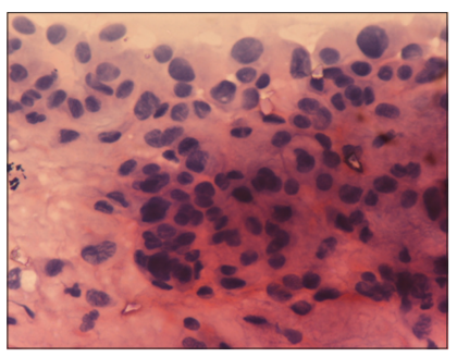
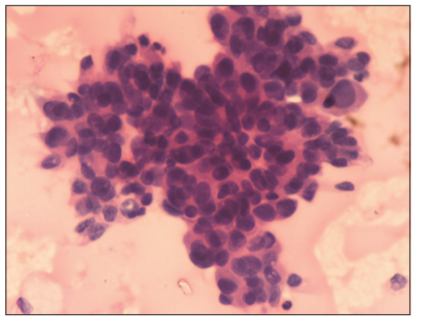
A 55-year-old male patient presented with complaints of swelling on the left side of the face for 22 years and pain in the same region for 4 months. Examination revealed a tender, slightly mobile, multinodular soft to firm swelling measuring 10 cm * 8 cm in the region of deep part of the parotid gland. The associated pain was sharp, intermittent, and radiating to the region above the ear. The patient gave a history of similar swelling 10 years back, which was operated on by some surgeon but without any documentary evidence. Intraoral findings did not give any significant clue. Fine-needle aspiration of the mass revealed cohesive clusters and singly arranged tumor cells with marked pleomorphism, with increased N/C ratio, hyperchromatic nucleus, and mild eosinophilic cytoplasm. Foci of keratinization could be appreciated in few tumor cells [Figures 1 and 2]. A cytological diagnosis suggestive of CxPA with squamoid differentiation was given. A left total parotidectomy was performed. The tumor was confined to the deep lobe of the parotid gland extending as deep as the deep parapharyngeal space. The tumor was deep to the facial nerve, with the nerve stretched downward and outward.


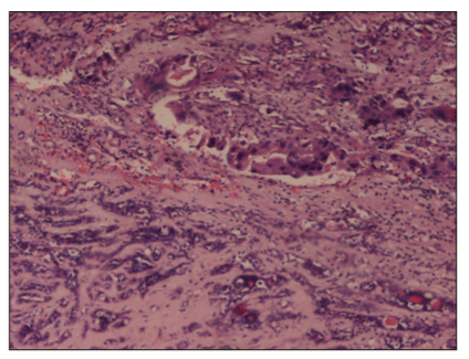
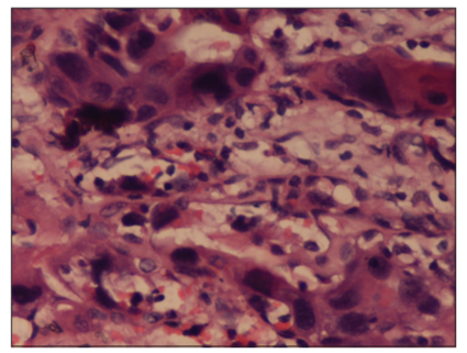
The lesion was grossly firm with infiltrative margins and areas of necrosis and hemorrhage. Microscopically, tissue section showed cluster of large, pleomorphic tumor cells with hyperchromatic nuclei, and well-outlined eosinophilic cytoplasm with foci of hemorrhage and necrosis, in a chondromyxoid background with benign epithelial and myoepithelial cells [Figure 3]. The malignant squamoid cells showed evidence of keratinization [Figure 4]. No capsular infiltration with the tumor cells was noted. A histological diagnosis of CxPA with squamoid differentiation was confirmed. Post-adjuvant chemotherapy in the form of Cisplatinum 50 mg * 6 cycles was administered to our patient. After 18 months of follow-up, our patient is doing well.


DISCUSSION
CxPA is seen in patients in the sixth to seventh decades of life[1]. Malignant transformation may occur up to 50 years after a pleomorphic adenoma is first diagnosed; the average period before malignant transformation is 20 years, as was evident in our case.[2,3].
CxPA is an infrequent aggressive malignancy with a high mortality and regional metastasis[2]. This type of tumor is difficult to diagnose, because the mixed tumor component is often small and overlooked, and the malignant component may be difficult to classify[2,3]. CxPAs are difficult to identify preoperatively, as they are more frequently localized in the deep lobe, in contrast to most pleomorphic adenomas, which tend to occur in the superficial lobe leading to low accuracy and sensitivity of FNAC[4]. However, we could give a suggestive diagnosis of CxPA on cytology, which was confirmed by the histopathological examination.
The most frequent histologic subtype is a high-grade adenocarcinoma (not otherwise specified)[5]. Our study revealed a rare case of squamoid differentiation in an existing pleomorphic adenoma. Histopathologically, clusters of malignant squamoid cells were noted in a background of chondromyxoid stroma, with foci of hemorrhage and necrosis.
Significant prognostic factors for CxPA include tumor stage, grade, age, proportion of carcinoma, extent of invasion, cervical metastasis, and proliferation index[3,6]. Our patient was a male aged 55 years which were similar to an average age of 56.3 years, as reported by Lewis et al.[7] In contrast, other reports have shown a female predominance[3,8]. It has been reported that if the carcinomatous component is contained within the benign mixed tumor capsule, as in the present case, the prognosis is excellent[9].
CxPA poses a challenge to diagnosis and treatment. Optimal diagnosis and treatment can be achieved only by close communication between the surgeon and pathologist, adequate surgery by an experienced oncologic surgeon, and assistance from a radiation oncologist[10]. The role of chemotherapy is quite promising in squamous cell carcinoma, as was seen in our case. After 18 months of follow-up post-chemotherapy, our patient is doing well.
We conclude that the best prevention of CXPA remains early and adequate removal of parotid neoplasms and prompt diagnosis and treatment of CXPA with squamoid differentiation can lead to complete cure of the patient.
REFERENCES
- Seifert G. Histopathology of malignant salivary gland tumours. Eur J Cancer 1992;28:49-56.
- Patil S, Gadbail AR, Chaudhary M. Carcinoma ex pleomorphic adenoma of parotid gland: Clinicopathological and immunohistochemical study of a case. Oral Surg 2009;2:182-7.
- Stodulski D, Rzepko R, Kowalska B, Stankiewicz C. Carcinoma ex pleomorphic adenoma of major salivary glands - A clinicopathologic review. Otolaryngol Pol 2007;61:687-93.
- Zbaren P, Zbaren S, Caversaccio MD, Stauffer E. Carcinoma ex pleomorphic adenoma: Diagnostic difficulty and outcome. Otolaryngol Head Neck Surg 2008;138:601-5.
- Olsen KD, Lewis JE. Carcinoma ex pleomorphic adenoma: A clinicopathologic review. Head Neck 2001;23:705-12.
- Lima RA, Tavares MR, Dias FL, Kligerman J, Nascimento MF, Barbosa MM, et al. Clinical prognostic factors in malignant parotid gland tumors. Otolaryngol Head Neck Surg 2005;133:702-8.
- Lewis JE, Olsen KD, Sebo TJ. Carcinoma ex pleomorphic adenoma: Pathologic analysis of 73 cases. Hum Pathol 2001;32:596-604.
- Brandwein M, Huvos AG, Dardick I, Thomas MJ, Theise ND. Carcinoma ex pleomorphic adenoma: A clinicopathologic review. Oral Surg Oral Med Oral Pathol Oral Radiol Endod 1996;21:23-4.
- Teo PM, Chan AT, Lee WY, Leung SF, Chan ES, Mok CO. Failure patterns and factors affecting prognosis of salivary gland carcinoma: Retrospective study. Hong Kong Med J 2000;6:29-36.
- Bell RB, Dierks EJ, Homer L, Potter BE. Management and outcome of patients with malignant salivary gland tumors. J Oral Maxillofac Surg 2005;63:917-28